NM_058216.2:c.145+1G>A is a canonical splice donor variant affecting the invariant +1 position of RAD51C intron 1, predicted to abolish normal splicing (SpliceAI delta 0.86). PVS1 is applied at very strong strength.1 Functional studies of c.145+1G>T, a different nucleotide substitution at the same canonical +1 position, demonstrated complete inactivation of the 5' splice site in a minigene splicing reporter assay and loss of normal RAD51C transcript expression in patient leukocytes (Meindl et al., 2010). PS3 is applied at supporting strength.2 This variant is extremely rare in population databases: gnomAD v2.1 AF = 3.98e-6 (1/251,282 alleles) and v4.1 AF = 6.20e-7 (1/1,614,194 alleles), with no homozygotes observed. PM2 is applied at moderate strength.3 This variant has been reported in ClinVar (Variation ID 484741) as Likely pathogenic by four clinical laboratories and Pathogenic by one clinical laboratory, with review status of criteria provided, single submitter.4
Final classification
Pathogenic
PVS1PS3PM2
RAD51C
c.145+1G>A
p.?
This variant
NM_058216.2:c.145+1G>A is a canonical splice donor variant affecting the invariant +1 position of RAD51C intron 1, predicted to abolish normal splicing (SpliceAI delta 0.86). PVS1 is applied at very strong strength.
ClinGen Hereditary Breast, Ovarian and Pancreatic Cancer Expert Panel Specifications to the ACMG/AMP Variant Interpretation Guidelines for RAD51C Version 1.0 v1.0 lacked a usable explicit final combination framework, so generic ACMG/AMP 2015 final-combination rules were applied as fallback; applied criteria: PVS1 very strong, PS3 supporting, PM2 moderate; combination = 1 very strong + 1 moderate + 1 supporting, which maps to Pathogenic.
Classification rationale
PVS1PS3PM2
Pathogenic
RAD51C c.145+1G>A
PVS1 + PS3 + PM2
→
Pathogenic
LYFE Sciences is an AI system, and it can make mistakes. Criteria
may be applied incorrectly, sources may be misread, and a confident-looking
classification can still be wrong. Double-check every criterion and
its underlying evidence before relying on any call.
Gene diagram
· NM_058216.2 · variants mapped to exon structure
RAD51C
NM_058216.2
Fetching transcript structure from UCSC…
Applied criteria · 3 applied · 17 assessed
MetEvidence satisfies this criterion.
Not metEvaluated against available evidence; threshold not reached.
Not assessedApplies in principle, but no evidence was found to evaluate it.
N/ADoesn't apply to this variant type.
Applied · 3
Strength
Supporting
Moderate
Strong
Very strong
PVS1
very strong
Pathogenic
NM_058216.2:c.145+1G>A is a canonical splice donor variant affecting the +1 position of intron 1, disrupting the invariant GT dinucleotide. RAD51C loss of function is an established disease mechanism for autosomal dominant hereditary breast and ovarian cancer and autosomal recessive Fanconi anemia. SpliceAI predicts a strong splice-altering effect (max delta score 0.86). The variant is predicted to abolish normal RAD51C transcript expression, consistent with experimental data showing complete inactivation of the 5' splice site when the same canonical position is disrupted by a different nucleotide substitution (c.145+1G>T evaluated in Meindl et al. 2010). PVS1 is applied at very strong strength under the generic ACMG/ClinGen SVI PVS1 framework (PMC6185798).
Canonical +1 splice donor variant disrupting invariant GT dinucleotideSpliceAI max delta score 0.86 predicts strong splice disruptionRAD51C loss of function is established germline disease mechanism
PS3
supporting
Pathogenic
Meindl et al. (2010, PMID:20400964) experimentally characterized c.145+1G>T, a different nucleotide substitution at the same canonical +1 splice donor position. RT-PCR of patient leukocytes showed reduced normal RAD51C-001 transcript and increased nonfunctional RAD51C-008 transcript. A minigene splicing reporter assay in HeLa cells demonstrated complete inactivation of the mutant 5' splice site. LOH of the wild-type allele was confirmed in tumor tissue. Although the exact variant (G>A) was not directly tested, both G>T and G>A disrupt the invariant GT dinucleotide at the canonical +1 position, and the mechanism of splice disruption is identical. Supporting strength is applied because the exact variant was not tested but functional data on the same position strongly supports a deleterious splice effect.
c.145+1G>T experimentally shown to inactivate 5' splice site by RT-PCR and minigene splicing reporter assayComplete loss of normal RAD51C-001 transcriptincreased nonfunctional RAD51C-008
PM2
moderate
Pathogenic
This variant is extremely rare in population databases. gnomAD v2.1 reports an allele frequency of 3.98e-6 (1/251,282 alleles, 0 homozygotes) and gnomAD v4.1 reports an allele frequency of 6.20e-7 (1/1,614,194 alleles, 0 homozygotes). Both are well below the PM2 threshold of <0.1%. The highest subpopulation frequency is in East Asian populations (v2.1: 5.44e-5; v4.1: 2.23e-5).
gnomAD v2.1: AF = 3.98e-6 (1/251282)0 homozygotes
Assessed · not applied
· 17 not met · 0 not assessed
Pathogenic
PS2
No de novo occurrence data is available for this variant.
PS4
No case-control study with statistical significance has been published for NM_058216.2:c.145+1G>A specifically.
PM1
The variant is a canonical splice donor site at the exon 1/intron 1 boundary.
PM6
No de novo occurrence has been reported for this variant.
PP1
No segregation data is available for NM_058216.2:c.145+1G>A.
PP3
While SpliceAI (max delta 0.86) and BayesDel (0.66) predict a deleterious effect, the splice prediction evidence is already captured by PVS1 for this canonical splice variant.
PP4
No patient phenotype or family history data is available for adjudication.
PP5
PP5 requires a reputable source (e.g., ClinGen expert panel, 3-star ClinVar review status) to have classified the variant as pathogenic.
Benign
BA1
The allele frequency in gnomAD is well below the BA1 threshold of >1%.
BS1
The allele frequency in gnomAD is well below the BS1 threshold of >0.3%.
BS2
No data is available showing this variant observed in healthy adults with full penetrance expected.
BS3
BS3 requires well-established in vitro or in vivo functional studies showing no damaging effect on protein function or splicing.
BS4
No segregation data is available for NM_058216.2:c.145+1G>A showing lack of co-segregation with disease.
BP2
No data available showing this variant observed in trans with a known pathogenic variant or in cis with a pathogenic variant in a recessive disorder.
BP4
BP4 requires multiple lines of computational evidence suggesting no impact on the gene or gene product.
BP5
No data available showing this variant found in a case with an alternative molecular basis for disease.
BP6
BP6 requires a reputable source (3-star ClinVar expert panel) to have classified the variant as benign or likely benign.
N/A · 8
PS1 · PM3 · PM4 · PM5 · PP2 · BP1 · BP3 · BP7
Research & evidence
Population frequency · supports pathogenic
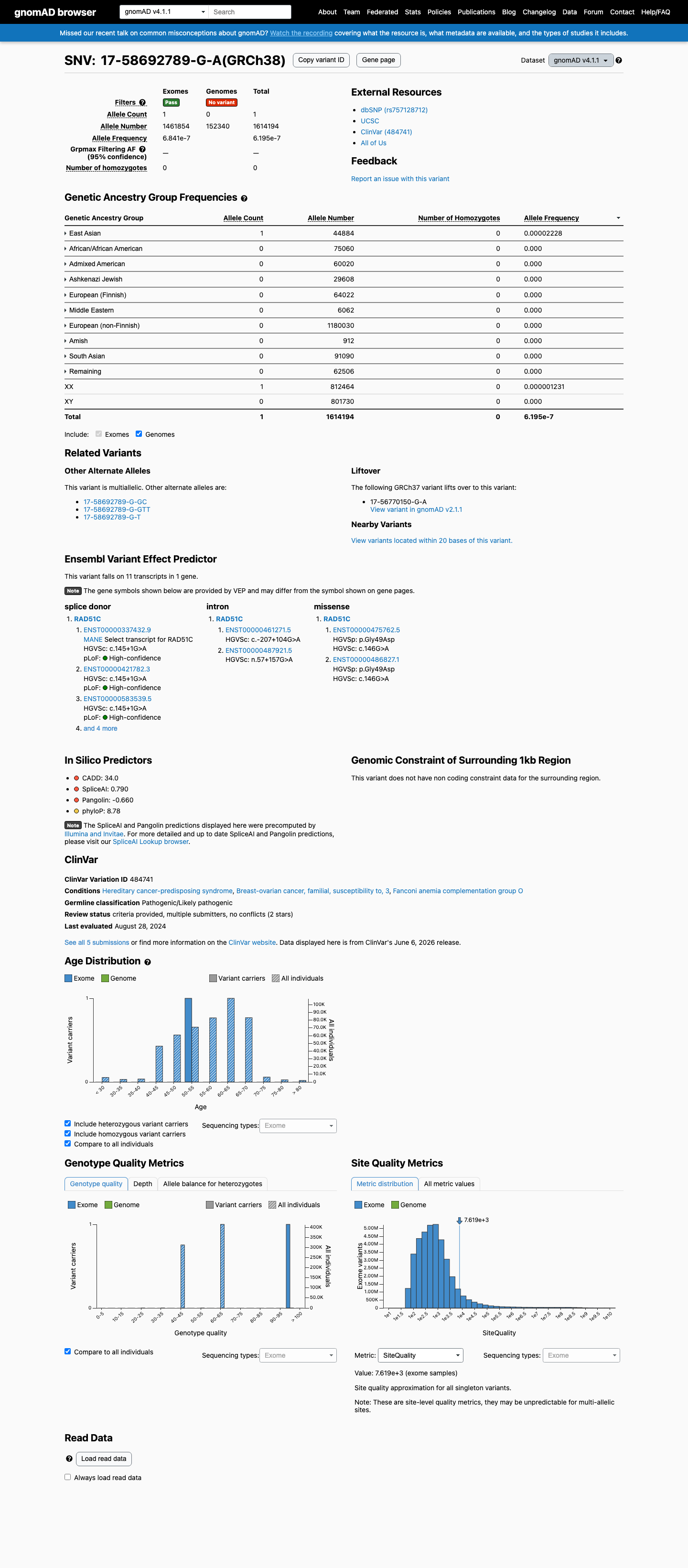
gnomAD v4.1
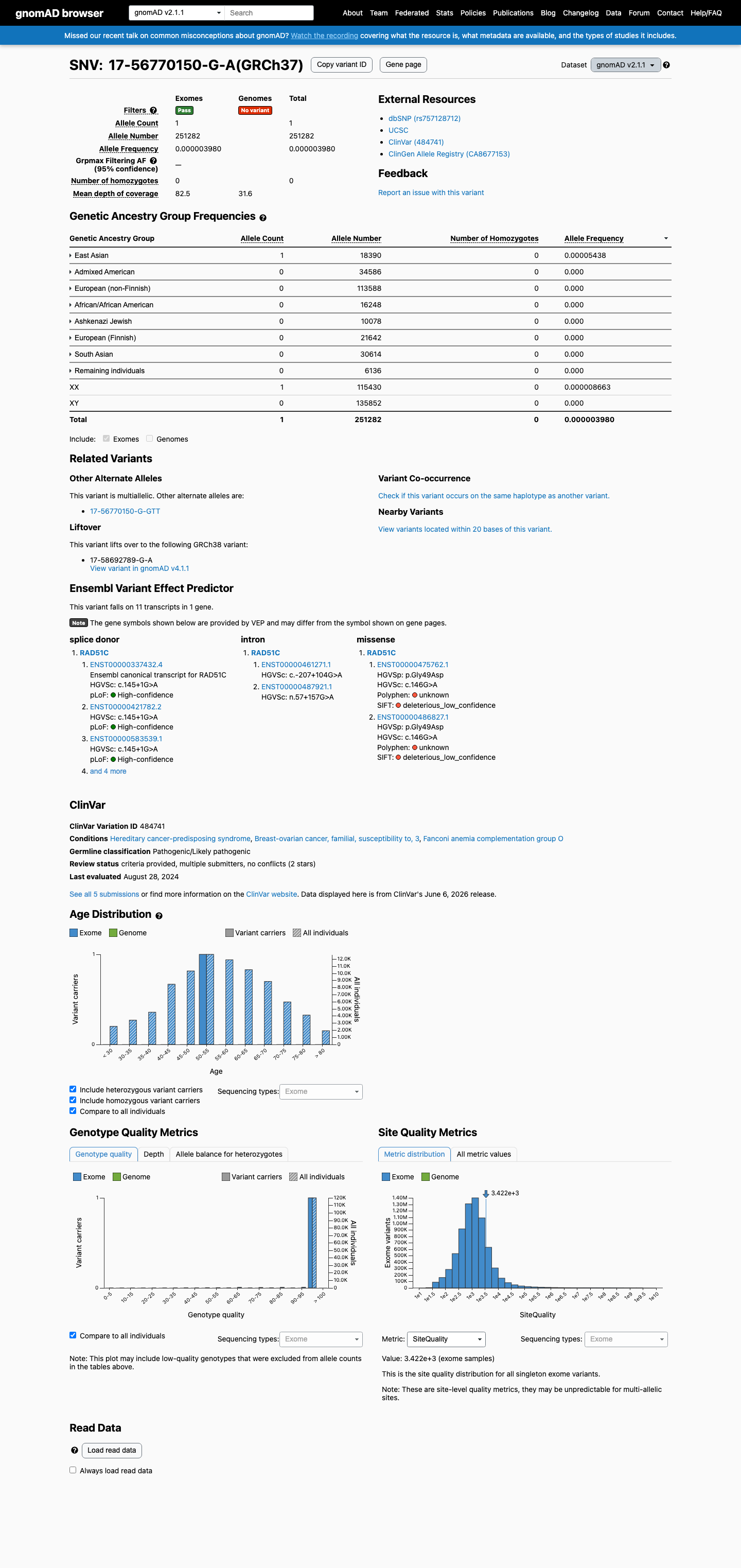
gnomAD v2.1
v4.1
This variant is present in gnomAD v4.1 (AF= 6.19504e-07; MAF= 0.00006%, 1/1614194 alleles, homozygotes = 0) and has highest observed frequency in the East Asian population (AF= 2.22797e-05; MAF= 0.00223%, 1/44884 alleles, homozygotes = 0).
v2.1
This variant is present in gnomAD v2.1 (AF= 3.97959e-06; MAF= 0.00040%, 1/251282 alleles, homozygotes = 0) and has highest observed frequency in the East Asian population (AF= 5.43774e-05; MAF= 0.00544%, 1/18390 alleles, homozygotes = 0).
🇨🇦 CA
Absent from gnomAD-Canada v1.0.
Allele frequency by ancestry
three datasets · side by side
gnomAD v4.1
6.2e-05%
· 1 / 1,614,194
0 hom
0 hom
East Asian 1 / 44,884 |
0.0022% |
+ 9 not observed (Remaining individuals, Admixed American, European (Finnish), Amish, Middle Eastern, South Asian, Ashkenazi Jewish, African/African American, European (non-Finnish))
gnomAD v2.1
0.0004%
· 1 / 251,282
0 hom
0 hom
East Asian 1 / 18,390 |
0.0054% |
+ 7 not observed (African/African American, Admixed American, Ashkenazi Jewish, European (Finnish), European (non-Finnish), Remaining individuals, South Asian)
gnomAD Canada 🇨🇦
Absent
· 0 / ?
0 hom
0 hom
Not observed in any ancestry group.
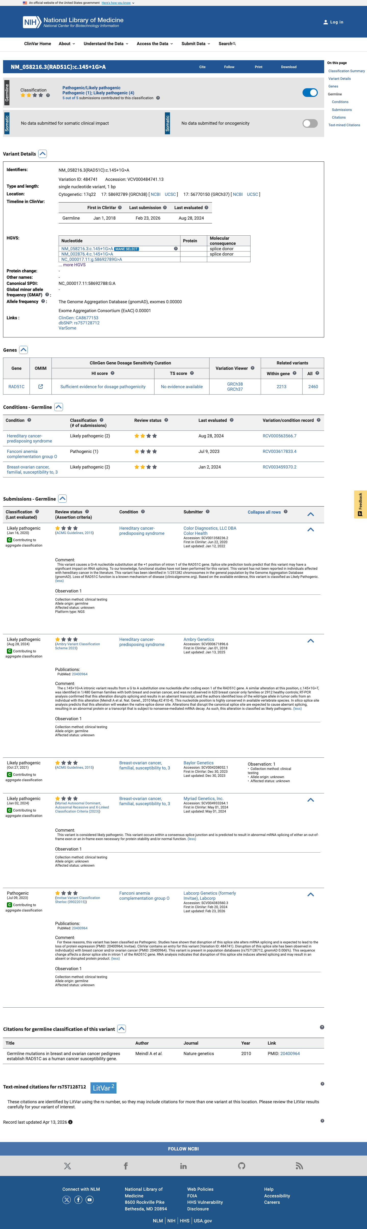
ClinVar
This variant has been reported in ClinVar as Likely pathogenic (4 clinical laboratories) and as Pathogenic (1 clinical laboratory). (ClinVarID = 484741)
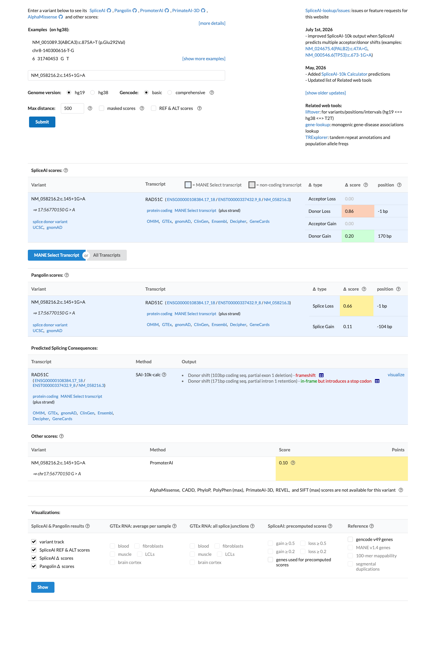
In silico
SpliceAI predicts possible splice impact for this variant (max delta score = 0.86). BayesDel score = 0.66.
Functional
No data
No calibrated functional assay or RNA evidence was identified for this variant.
COSMIC
Somatic evidence
COSMIC
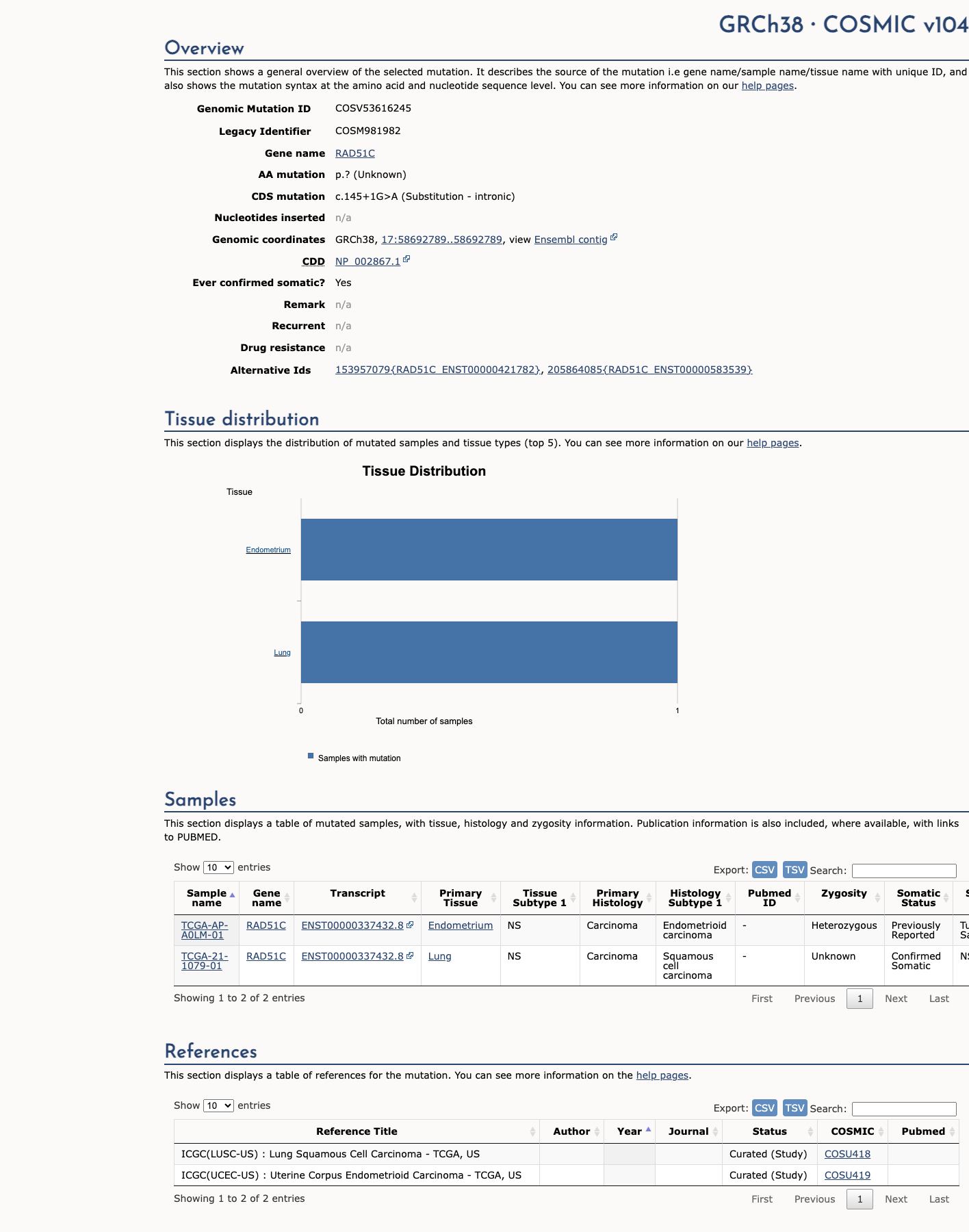
This variant has previously been reported in somatic cancers (COSMIC; COSV53616245, n = 2 times).
Hotspots
This variant does not lie in a statistically significant cancer hotspot.
Literature · how each cited paper was used
1papers cited
Each card is an audit: what was searched, what was found, whether it names the variant, which criteria it fed, and why. 3 further PMIDs triaged but not cited — see Sources & references.
Rule & framework references · cited for criterion definitions, not variant evidence
20400964 ↗
Germline mutations in breast and ovarian cancer pedigrees establish RAD51C as a human cancer susceptibility gene.
Sources & reference links
Triaged references · 3 PMIDs not cited in assessment
25741868 ↗
Standards and guidelines for the interpretation of sequence variants: a joint consensus recommendation of the American College of Medical Genetics and Genomics and the Association for Molecular Pathology.
CLINVAR
25394175 ↗
A practice guideline from the American College of Medical Genetics and Genomics and the National Society of Genetic Counselors: referral indications for cancer predisposition assessment.
CLINVAR