NM_033632.3:c.1436G>T (p.Arg479Leu) is a missense variant in exon 10 of FBXW7, located at residue 479 within the WD40 substrate recognition domain. This variant is absent from gnomAD v2.1, v4.1, and gnomAD-Canada population databases, meeting PM2 at supporting strength.1 The variant lies at a statistically significant mutational hotspot (cancerhotspots.org) within the WD40 domain, a well-characterized functional domain critical for FBXW7 substrate recognition. Mutations at residue 479 have been shown to disrupt FBXW7-substrate interactions across multiple studies, meeting PM1 at moderate strength.2 Functional studies directly testing the R479L substitution in a co-immunoprecipitation assay (293T cells) demonstrated complete loss of FBXW7 interaction with its substrate DAB2IP, confirming disruption of substrate recognition function. This meets PS3 at moderate strength.3 Multiple in silico tools support a deleterious effect: REVEL score 0.583 and SpliceAI max delta score 0.33 (acceptor loss prediction). This meets PP3 at supporting strength.4 The variant has been reported in ClinVar (Variation ID: 4530538) as Tier I - Strong with a single submitter (1-star review status). It has been observed in somatic cancers (COSMIC COSV55899054, 18 counts) but lacks independent germline proband observations. These data are insufficient for PP5 or PS4.5
FBXW7
Final classification
Likely Pathogenic
FBXW7 c.1436G>T · p.Arg479Leu
FBXW7
NM_033632.3:c.1436G>T (p.Arg479Leu) is a missense variant in exon 10 of FBXW7, located at residue 479 within the WD40 substrate recognition domain.
gene-specific framework lacked a usable explicit final combination framework, so generic ACMG/AMP 2015 final-combination rules were applied as fallback; applied criteria: PS3 moderate, PM1 moderate, PM2 supporting, PP3 supporting; combination = 2 moderate + 2 supporting, which maps to Likely Pathogenic.
Classification rationale
PS3PM1PM2PP3
Likely Pathogenic
FBXW7 c.1436G>T
PS3 + PM1 + PM2 + PP3
→
Likely Pathogenic
Gene diagram
· NM_033632.3 · variants mapped to exon structure
FBXW7
NM_033632.3
Fetching transcript structure from UCSC…
Applied criteria · 4 applied · 19 assessed
Applied · 4
Strength
Supporting
Moderate
Strong
Very strong
✓
PS3
moderate
Pathogenic
The R479L substitution was directly tested in a co-immunoprecipitation assay in 293T cells and demonstrated complete loss of FBXW7 interaction with its substrate DAB2IP, confirming disruption of substrate recognition function. This is a single study with direct variant testing using an indirect reporter-type assay, supporting moderate strength.
PMID:24912918: 293T co-immunoprecipitation assay showing R479L abolishes FBXW7-DAB2IP interactionconfirming loss of substrate recognition.
✓
PM1
moderate
Pathogenic
The variant is located at residue Arg479 within the WD40 substrate recognition domain of FBXW7, a well-characterized critical functional domain. This residue is a statistically significant mutational hotspot per cancerhotspots.org. Functional studies (PMID:24912918, PMID:17646408, PMID:17646409) confirm that mutations at this position disrupt FBXW7 substrate binding. The variant lies in the WD40 repeat domain that mediates substrate recognition, and no benign variation is observed at this position in population databases.
Cancerhotspots.org: statistically significant mutational hotspot at residue 479.PMID:24912918: R479L disrupts FBXW7-DAB2IP interaction.PMID:17646408: R479Q mutation in T-ALL disrupts FBXW7-NOTCH1 interaction.
✓
PM2
supporting
Pathogenic
This variant is absent from gnomAD v2.1 (exomes), gnomAD v4.1 (exomes), and gnomAD-Canada v1.0. Allele frequency is below the 0.1% PM2 threshold for rare variant support in the generic ACMG framework.
Absent from gnomAD v2.1.Absent from gnomAD v4.1.Absent from gnomAD-Canada v1.0.
✓
PP3
supporting
Pathogenic
Multiple lines of computational evidence support a deleterious effect: REVEL score of 0.583 (above the 0.5 threshold for pathogenicity) and SpliceAI predicts a possible splice alteration with a max delta score of 0.33 (acceptor loss). The variant also lies at a statistically significant mutational hotspot. BayesDel score is low at 0.125 but does not outweigh the preponderance of evidence from REVEL and SpliceAI.
REVEL: 0.583 (deleteriousabove 0.5 threshold).SpliceAI: max delta 0.33 (acceptor loss Δ = 0.33
Assessed · not applied
Pathogenic
PS1
PS1 requires a different nucleotide change at the same codon producing the same amino acid change (p.Arg479Leu) that has been established as pathogenic.
PS2
PS2 requires a de novo observation with confirmed maternity and paternity.
PS4
PS4 requires statistically significant enrichment in affected individuals or a substantial number of independent proband observations.
PM5
PM5 requires a different pathogenic missense variant at the same amino acid residue (Arg479).
PM6
PM6 requires a de novo observation with confirmed maternity and paternity.
PP1
PP1 requires cosegregation of the variant with disease in multiple affected family members.
PP2
PP2 applies when a gene has a low rate of benign missense variation and missense variants are a known disease mechanism.
PP4
PP4 requires the variant to be found in a patient with a phenotype highly specific for FBXW7-related disease.
PP5
PP5 requires a reputable source (≥3-star ClinVar expert panel) to have classified the variant as pathogenic.
Benign
BA1
BA1 requires an allele frequency >1% in gnomAD.
BS1
BS1 requires an allele frequency >0.3% in gnomAD.
BS2
BS2 requires observation in a healthy adult at a frequency inconsistent with a fully penetrant disorder.
BS3
BS3 requires well-established functional studies showing no damaging effect.
BS4
BS4 requires lack of segregation in affected family members.
BP1
BP1 applies to missense variants in genes where only truncating variants cause disease.
BP2
BP2 requires observation in trans with a pathogenic variant for a fully penetrant dominant disorder.
BP4
BP4 requires multiple lines of computational evidence suggesting no impact on gene product.
BP5
BP5 requires the variant to be found in a case with an alternate molecular basis for disease.
BP6
BP6 requires a reputable source (≥3-star ClinVar EP) to have classified the variant as benign.
N/A · 5
PVS1 · PM3 · PM4 · BP3 · BP7
Research & evidence
Population frequency · supports pathogenic
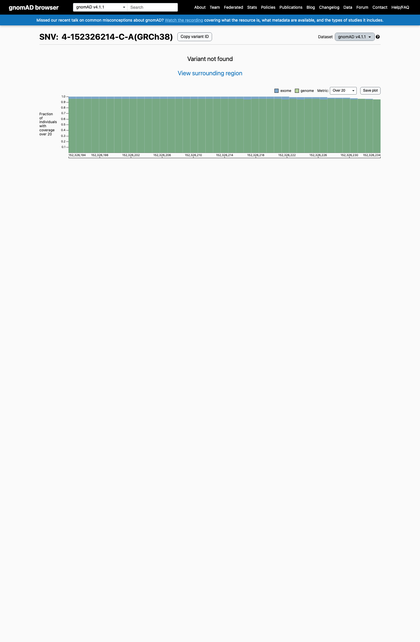
gnomAD v4.1
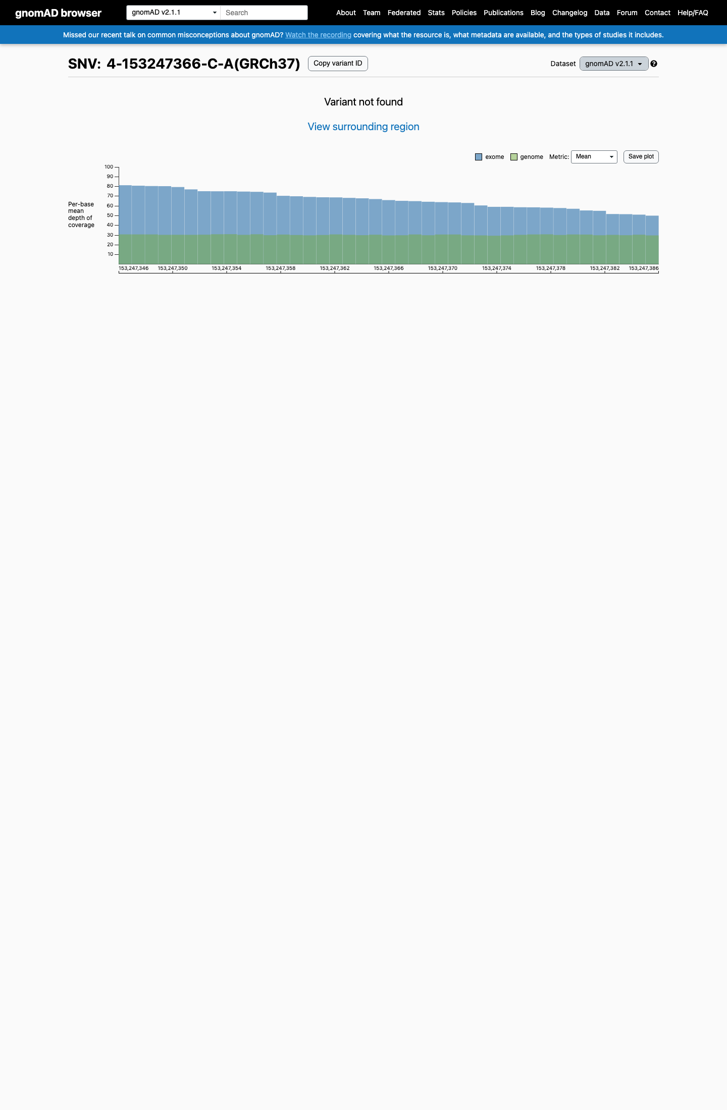
gnomAD v2.1
v4.1
Absent from gnomAD v4.1.
v2.1
Absent from gnomAD v2.1.
🇨🇦 CA
Absent from gnomAD-Canada v1.0.
Allele frequency by ancestry
three datasets · side by side
gnomAD v4.1
Absent
· 0 / ?
0 hom
0 hom
Not observed in any ancestry group.
gnomAD v2.1
Absent
· 0 / ?
0 hom
0 hom
Not observed in any ancestry group.
gnomAD Canada 🇨🇦
Absent
· 0 / ?
0 hom
0 hom
Not observed in any ancestry group.
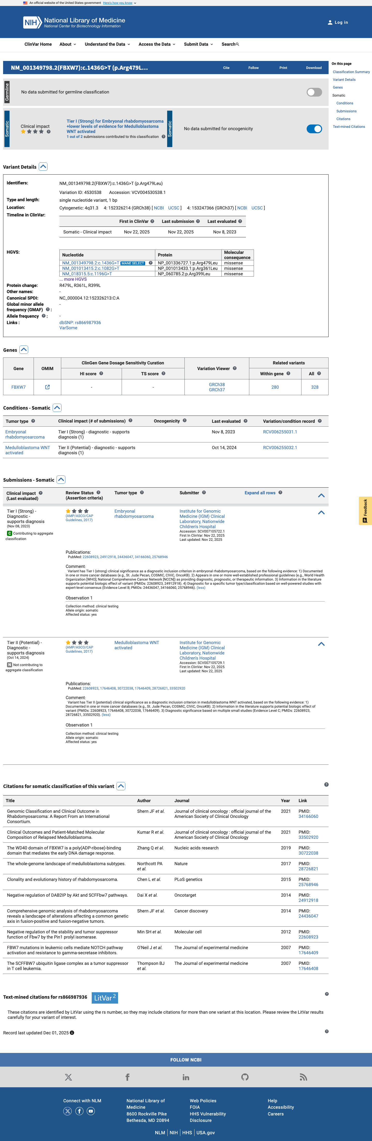
ClinVar
This variant has been reported in ClinVar but submission details could not be extracted. (ClinVarID = 4530538)
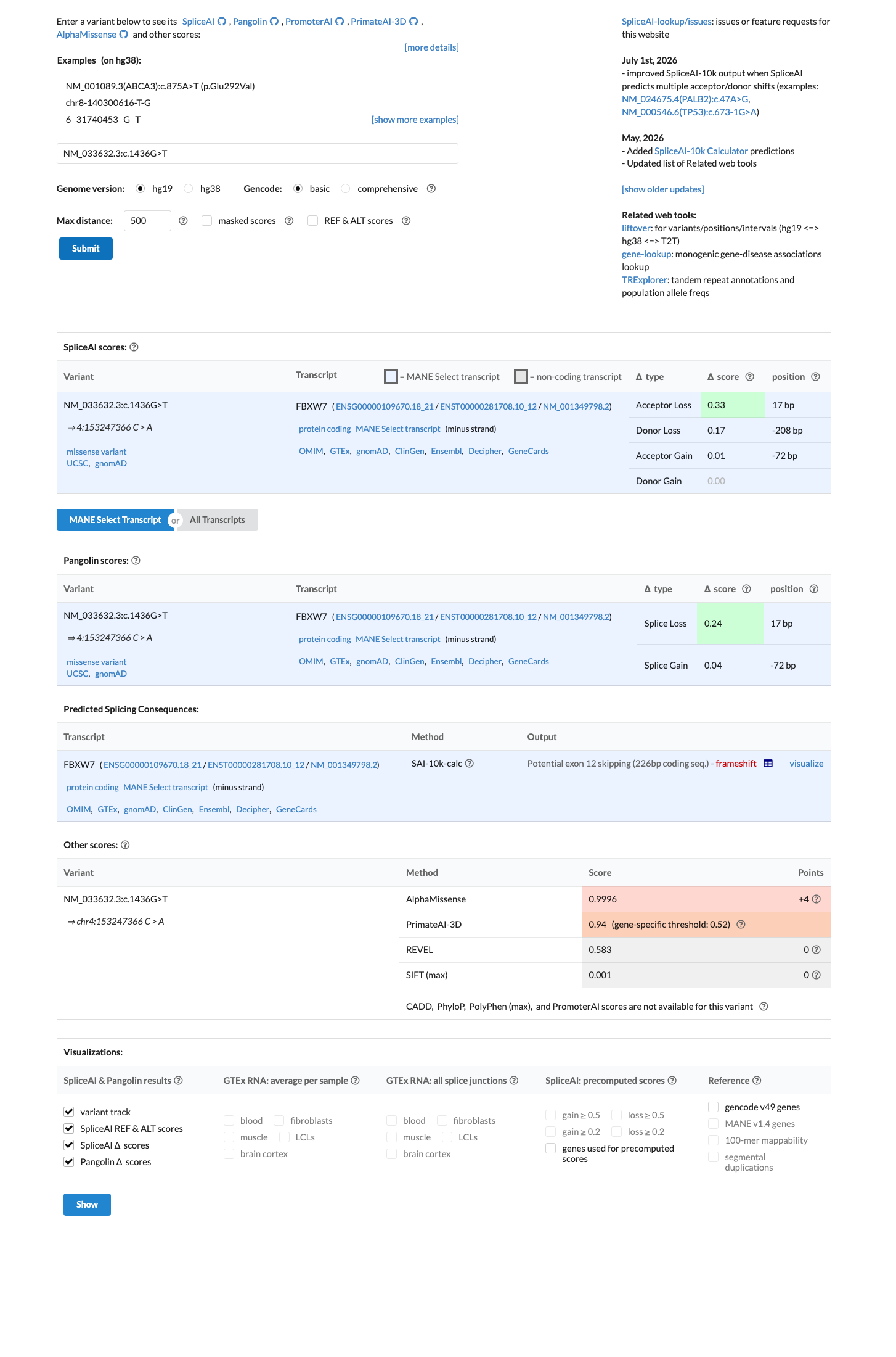
In silico
SpliceAI predicts possible splice impact for this variant (max delta score = 0.33). REVEL score = 0.583. BayesDel score = 0.124658.
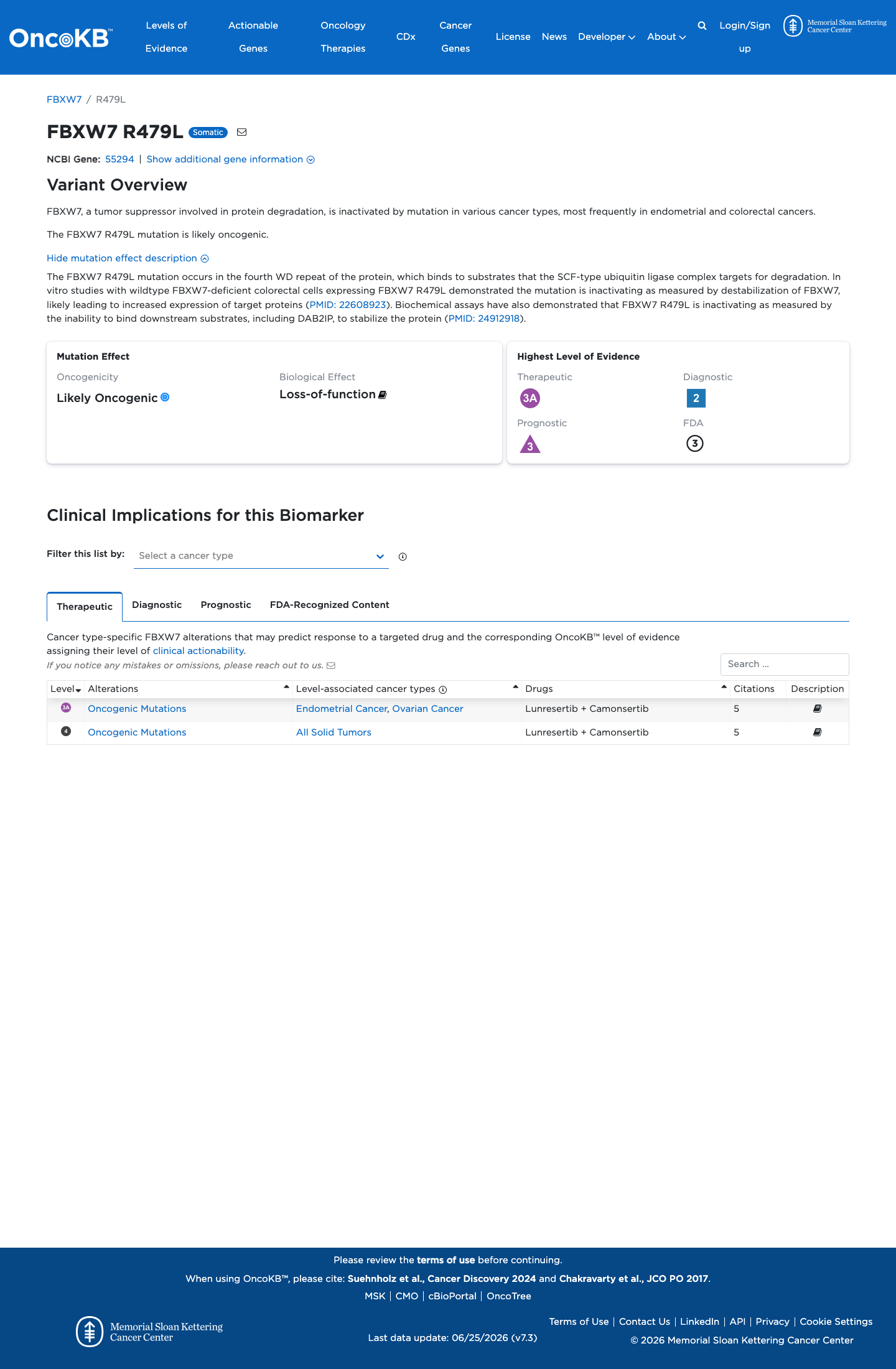
Functional
Likely Oncogenic
OncoKB identified variant-specific curated literature and context relevant to functional review; biological-effect context: Loss-of-function; curated oncogenicity label: Likely Oncogenic.
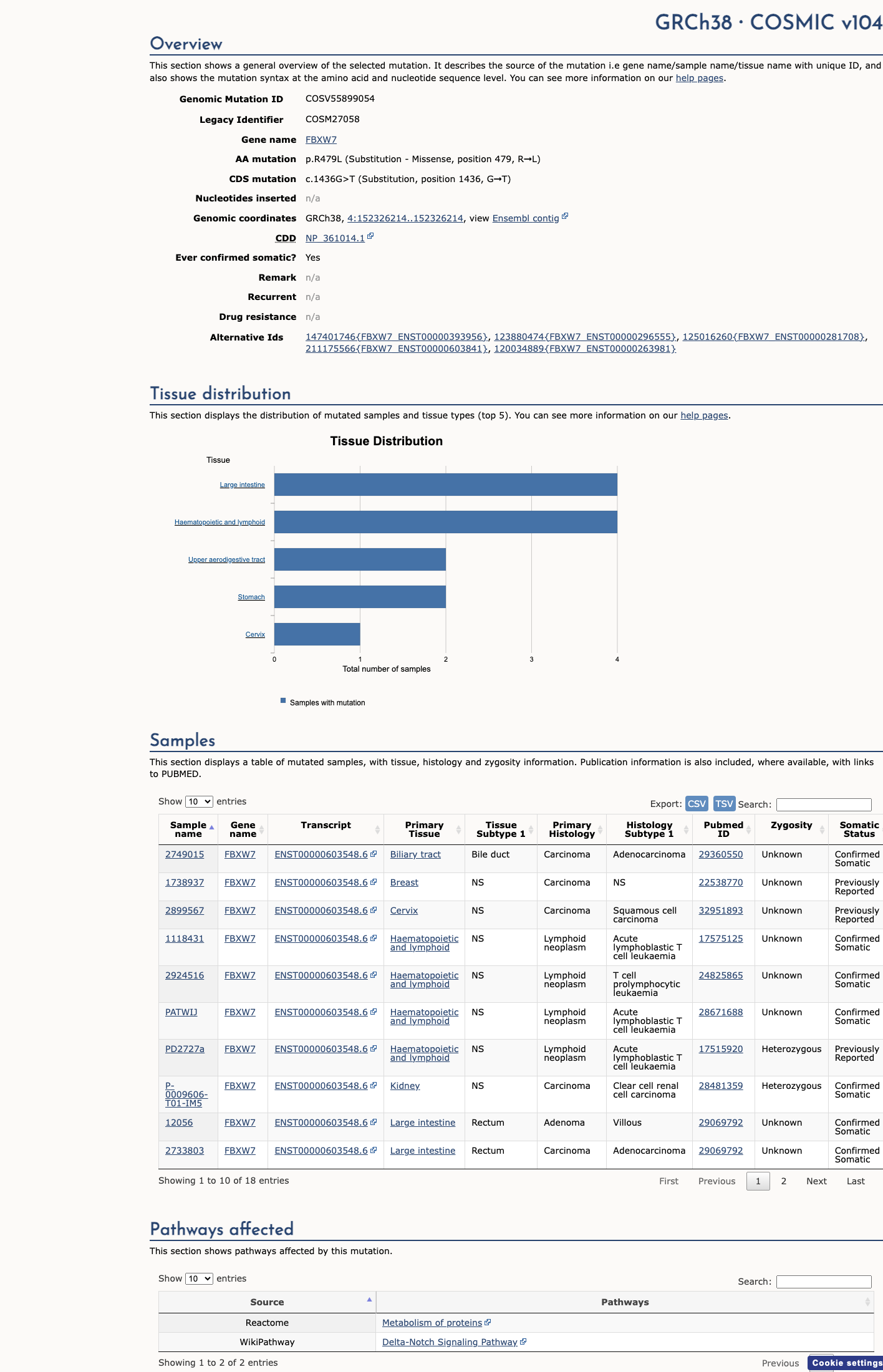
COSMIC

Cancer hotspots
Somatic evidence
Hotspot
COSMIC
This variant lies in a statistically significant hotspot. This variant has previously been reported in somatic cancers (COSMIC; COSV55899054, n = 18 times).
Hotspots
This variant lies in a statistically significant hotspot.
Literature · how each cited paper was used
3papers cited
Each card is an audit: what was searched, what was found, whether it names the variant, which criteria it fed, and why. 3 further PMIDs triaged but not cited — see Sources & References.
The SCFFBW7 ubiquitin ligase complex as a tumor suppressor in T cell leukemia.
Searched
R479LArg479Leuc.1436G>T1436R479
Found
Reports FBW7 mutations in T-ALL cell lines and primary samples, identifying R465H, R479Q, and R505C as recurrent mutations in the WD40 domain that disrupt NOTCH1 binding. The variant R479L was not studied; the paper examined R479Q (c.1585G>A), a different amino acid substitution at the same codon.
Variant
◇ Residue / gene-level — variant not named
Applied to
→PM1 supports · met
Why
R479Q (not R479L) was tested; cited in PM1 to support functional importance of residue 479 in the WD40 domain, but does not provide direct evidence for the specific variant under assessment.
we replaced the arginine at position 465, 479, or 505 with cysteine (DN FBW7 R465C and DN FBW7 R505C) or glutamine (DN FBW7 R479Q) and tested their abilities to interact with N1-IC
Location Results; Table of T-ALL mutations; Figure panels showing DN FBW7 mutants · Context Co-immunoprecipitation, T-ALL cell lines and 293T cells · full text
FBW7 mutations in leukemic cells mediate NOTCH pathway activation and resistance to gamma-secretase inhibitors.
Searched
R479LArg479Leuc.1436G>T1436R479
Found
Reports that FBW7 mutations in T-ALL cell lines (including R465H, R479Q, and R505C) mediate NOTCH pathway activation and resistance to gamma-secretase inhibitors. The R479Q mutant showed defective NOTCH binding with modest residual interaction. The variant R479L was not studied.
Variant
◇ Residue / gene-level — variant not named
Applied to
→PM1 supports · met
Why
R479Q (not R479L) was tested; cited in PM1 to support functional importance of residue 479 in the WD40 domain, but does not provide direct evidence for the specific variant under assessment.
the mutants were defective in NOTCH binding, although FBW7R479Q showed a modest amount of residual binding
Location Results, Figure 6A · Context Co-immunoprecipitation, T-ALL cell lines · full text
Negative regulation of DAB2IP by Akt and SCFFbw7 pathways.
Searched
R479LArg479Leuc.1436G>T1436
Found
Three T-ALL-associated mutations in FBXW7 (R465H, R479L, R505C) were tested in a co-immunoprecipitation assay and shown to abolish interaction with the substrate DAB2IP, confirming loss of substrate recognition function.
Variant
✓ Names this variant — characterised directly
Applied to
→PM1 supports · met
→PS3 supports · met
Why
Variant-specific functional data confirmed loss of substrate interaction; referenced in PS3 (moderate) and PM1 assessments.
Consistent with DAB2IP being a substrate of Fbw7, we found that three T-ALL associated mutations in Fbw7 (R465H, R479L, and R505C) no longer interacted of Fbw7 with DAB2IP (Figure 4B), providing further evidence that DAB2IP interaction with Fbw7 is through the substrate recognition domain of Fbw7.
Location Results, Figure 4B · Context Co-immunoprecipitation assay, 293T cells, FLAG-DAB2IP and HA-FBW7 constructs · full text
Sources & reference links
Triaged references · 3 PMIDs not cited in assessment
22608923 ↗
Negative regulation of the stability and tumor suppressor function of Fbw7 by the Pin1 prolyl isomerase.
ONCOKB
27993330 ↗
Standards and Guidelines for the Interpretation and Reporting of Sequence Variants in Cancer: A Joint Consensus Recommendation of the Association for Molecular Pathology, American Society of Clinical Oncology, and College of American Pathologists.
CLINVAR