This variant was interpreted by pipeline
7.1.0.
The current version is
8.0.0.
Re-running queues a fresh interpretation; the result replaces this page when complete.
Re-runs are not free. Blank = draws on today's public interpretation allowance.
With a code = uses 1 of that code's uses.
FANCD2 encodes a DNA repair protein that works together with its partner FANCI to coordinate the cellular response to DNA damage and maintain genome stability. Inherited mutations in FANCD2 cause Fanconi anemia, a rare disorder characterized by congenital defects, bone marrow failure, and an elevated risk of cancer. FANCD2 acts as a tumor suppressor: loss of its function impairs DNA repair and can promote cancer development, and altered FANCD2 activity has been observed in several tumor types, including leukemias, breast cancer, melanoma, and colorectal cancer.
This variant
FANCD2 loss-of-function causes autosomal recessive Fanconi anemia and predisposes to cancer, so a canonical splice-site change with near-certain predicted splice impact (SpliceAI max delta 0.99) is biologically concerning. However, this variant remains a VUS: only its absence from population databases was established, and no functional, clinical, or family data were available to confirm a disease-causing effect.
Transcript
NM_033084.4
HGVS · transcript:coding
NM_033084.4:c.3560+2T>A
GRCh38
chr3:10088544 T>A
GRCh37
chr3:10130228 T>A
Variant of Uncertain Significance: only PM2 (supporting) was met — the variant is absent (AF 0) from gnomAD — and one supporting criterion meets no Pathogenic, Likely Pathogenic, Benign, or Likely Benign threshold.
Classification rationale
PM2VUS
FANCD2 c.3560+2T>Acanonical splice
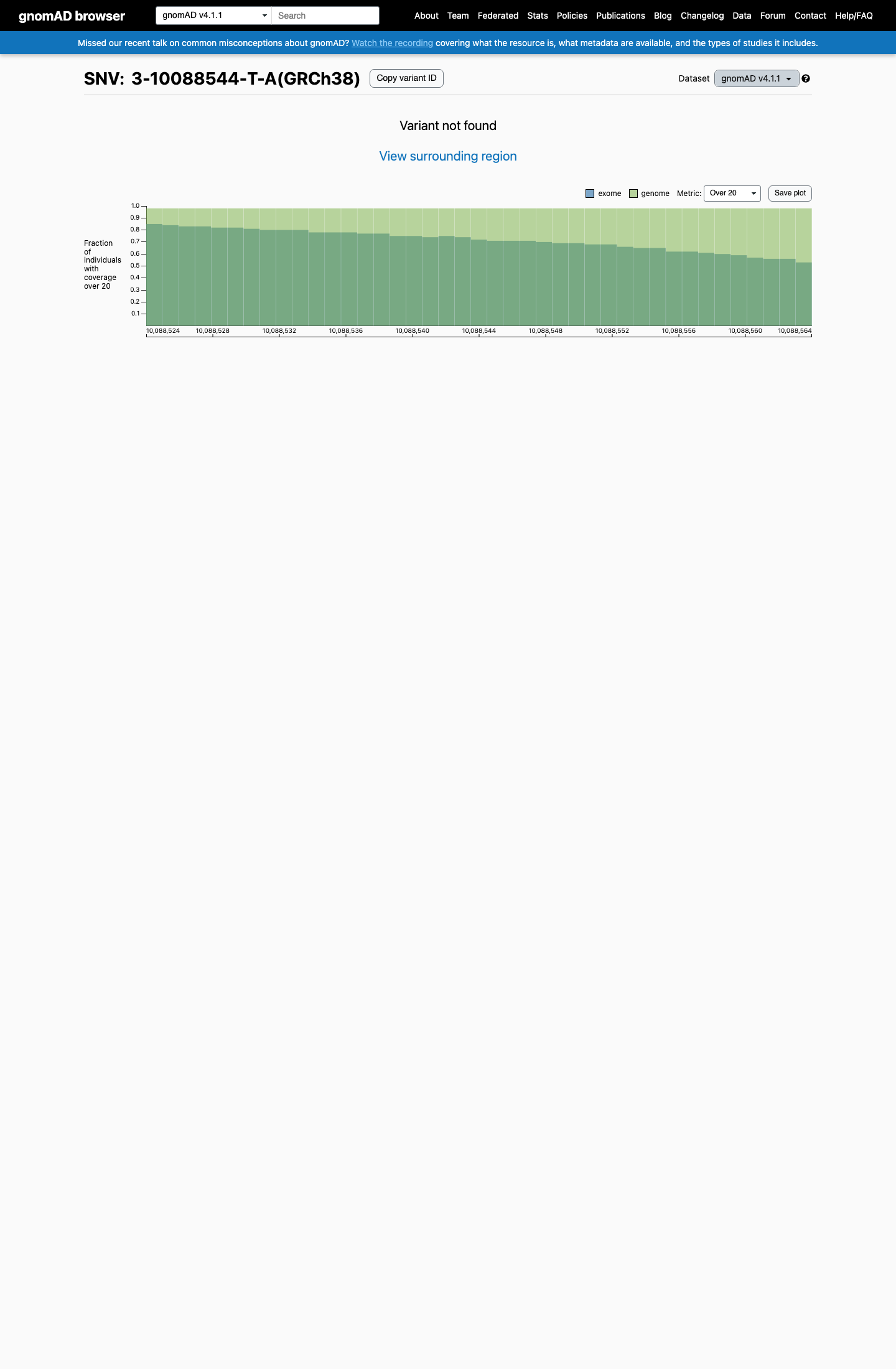
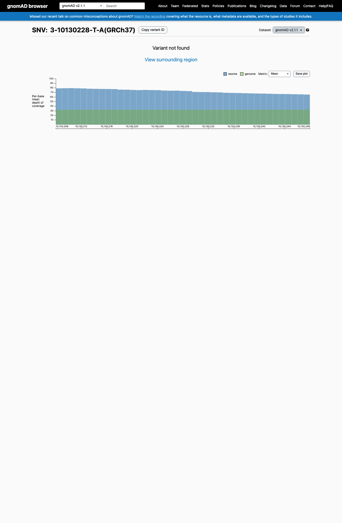
PM2 (Supporting): variant absent (AF 0) from gnomAD v2.1, v4.1, and gnomAD-Canada — below the <0.1% recessive-disorder threshold. Overall classification: VUS — a single supporting criterion meets no Pathogenic, Likely Pathogenic, Benign, or Likely Benign combination threshold under the generic ACMG/AMP 2015 framework.
PM2→VUS
LYFE Sciences is an AI system, and it can make mistakes. Criteria
may be applied incorrectly, sources may be misread, and a confident-looking
classification can still be wrong. Double-check every criterion and
its underlying evidence before relying on any call.
Gene diagram
· NM_033084.4 · variants mapped to exon structure
FANCD2NM_033084.4
Fetching transcript structure from UCSC…
Exons
—
Transcript span
—
Strand
—
Variants mapped
—
Source
UCSC ncbiRefSeqCurated
All variants in FANCD2—click a row to locate it on the plot · use the link column to open its page
Protein
Location
Classification
Link
Applied criteria · 1 applied · 19 assessed
MetEvidence satisfies this criterion.
Not metEvaluated against available evidence; threshold not reached.
Not assessedApplies in principle, but no evidence was found to evaluate it.
N/ADoesn't apply to this variant type.
Applied · 1
Strength Supporting Moderate Strong Very strong
✓
PM2supportingPathogenic
Met (supporting): variant is absent (AF 0) from gnomAD v2.1, v4.1, and gnomAD-Canada, below the <0.1% PM2 threshold.
Assessed · not applied
· 7 not met · 12 not assessed
Pathogenic
PVS1Not assessed: insufficient evidence was available to evaluate whether this splice variant causes loss of function.
PS2Not assessed: no proband or parental-testing data were available to confirm or exclude a de novo origin.
PS3Not assessed: no functional assay evidence was available; the only relevant datum was an in silico splice prediction, not a functional assay.
PS4Not assessed: no case-control or cohort data reported this variant — it is absent from ClinVar, gnomAD, and the literature.
PM3Not assessed: no proband, second-allele, or phase data were available to test for a pathogenic trans counterpart.
PM6Not assessed: no proband or parental testing data were available to evaluate an assumed de novo occurrence.
PP1Not assessed: no pedigree or family segregation data were available.
PP3Not met: SpliceAI max delta 0.99 predicts near-certain splice impact, but this same prediction is counted under PVS1 and cannot be double-counted as PP3.
PP4Not assessed: no proband phenotype or family history data were available to assess phenotype specificity.
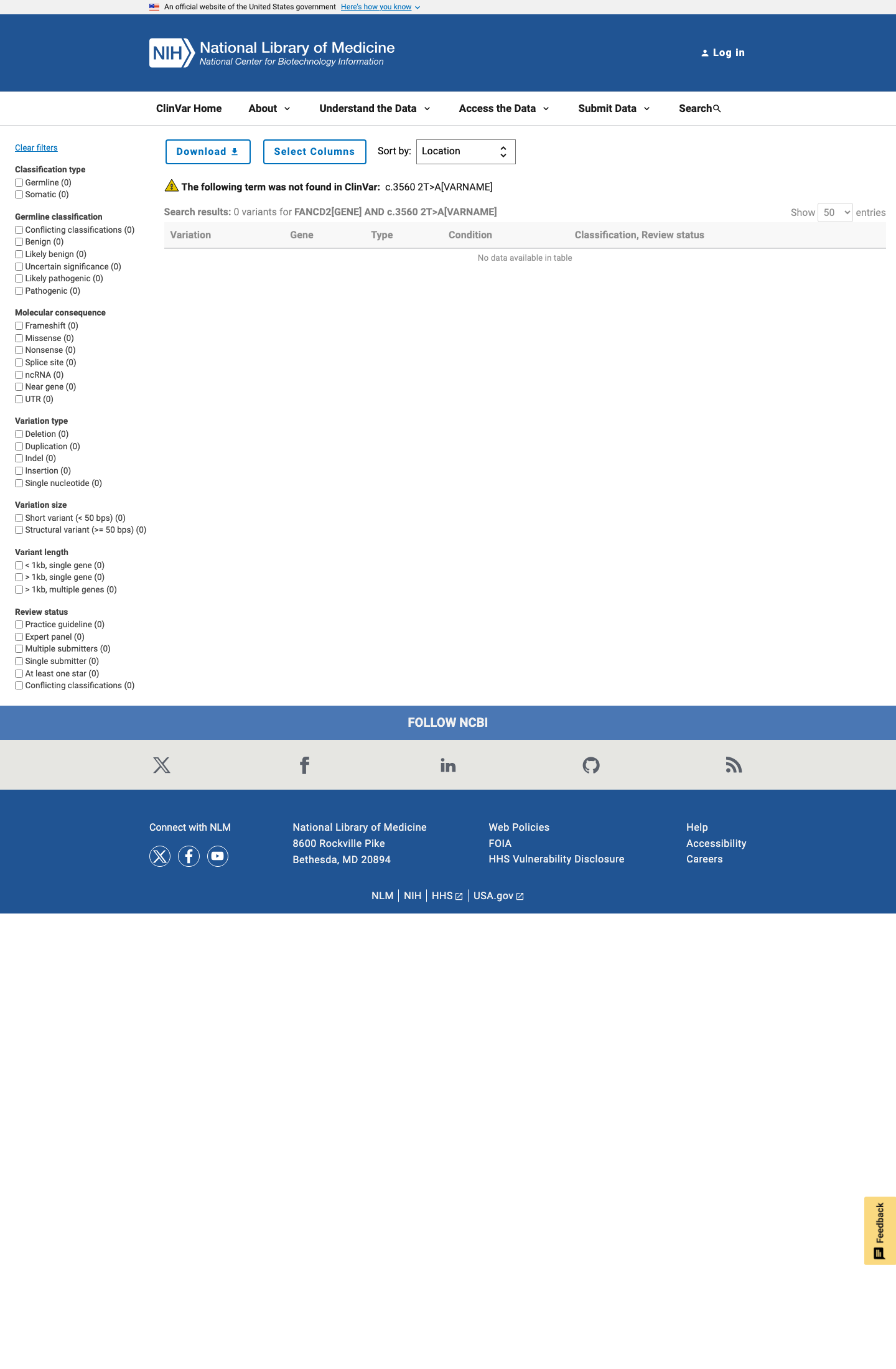
PP5Not met: ClinVar has no record for this variant, so no expert-panel Pathogenic or Likely pathogenic classification exists to support PP5.
Benign
BA1Not met: variant is absent (AF 0) from population databases, far below the >1% BA1 threshold.
BS1Not met: variant is absent (AF 0), far below the >0.3% BS1 threshold expected for this very rare recessive disorder.
BS2Not met: no homozygous observations exist — the variant is absent from all queried population databases, and biallelic loss causes severe early-onset disease.
BS3Not assessed: no well-established functional study demonstrating a benign effect was available for this variant.
BS4Not assessed: no family segregation testing was performed, so lack of segregation could not be observed.
BP2Not assessed: no second variant or phase-resolved observation was available to evaluate a cis/trans configuration.
BP4Not met: SpliceAI max delta 0.99 predicts near-certain splice impact, the opposite of the no-impact evidence BP4 requires.
BP5Not assessed: no proband genotype or diagnostic data were available to evaluate an alternative molecular cause.
BP6Not met: ClinVar has no record for this variant, so no expert-panel Benign or Likely benign classification exists to support BP6.